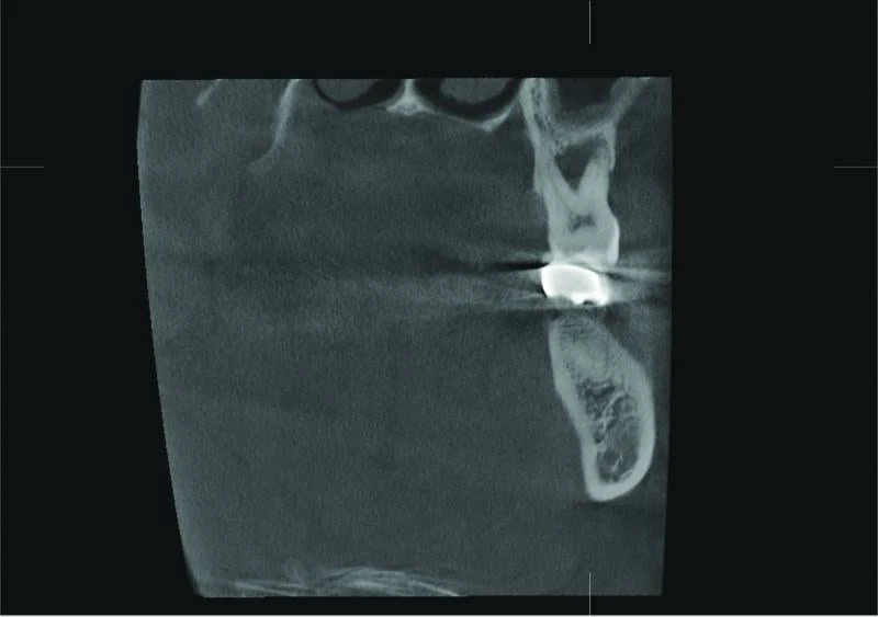
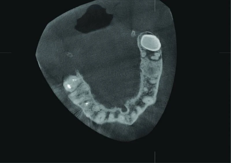
Images taken with Veraviewepocs 3D R100
Findings:
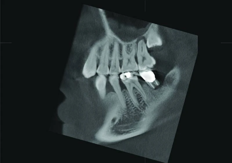
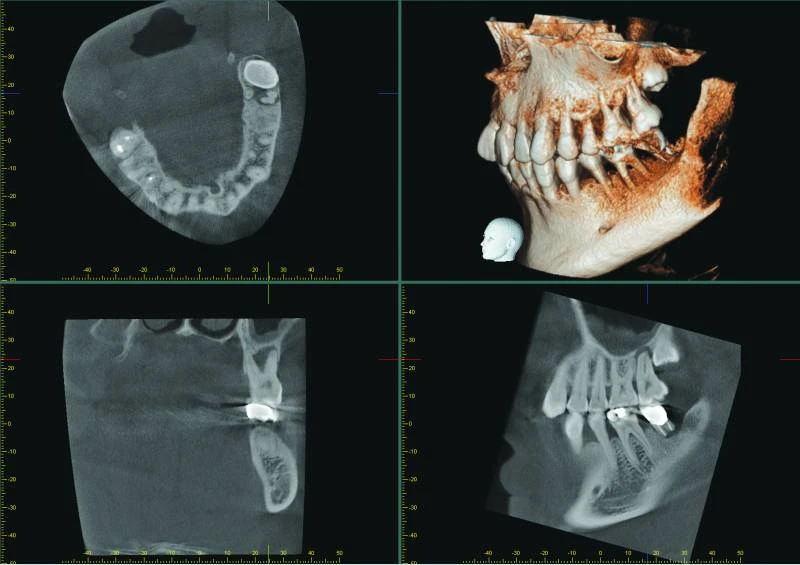
The patient presented with pain in the left upper jaw. A 3D x-ray was taken with Veraviewepocs 3D R100. Based on the x-ray it was determined that tooth 28 really was impacted and that tooth 27 was causing problems, too.
The axial view allowed the conclusion that there was substantial bone loss near the apex of tooth 27 as well as damage to the sinus floor and pachymenia.
Bài đăng lần đầu ngày: 27 Tháng Năm, 2018 @ 5:31 chiều