Images taken with Veraviewepocs 3D R100
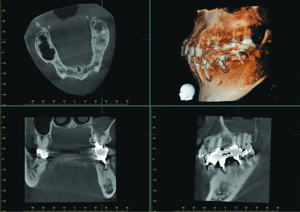
The patient presented with an unclear situation around tooth 26, which had undergone endodontic treatment before. Conventional 2D X-rays did not allow a conclusive diagnosis; therefore, a 3D scan was performed with Veraviewepocs 3D R100. The sagittal and coronal view showed that the endodontic treatment had not been successful and that there were apical defects on the buccal and palatal root.
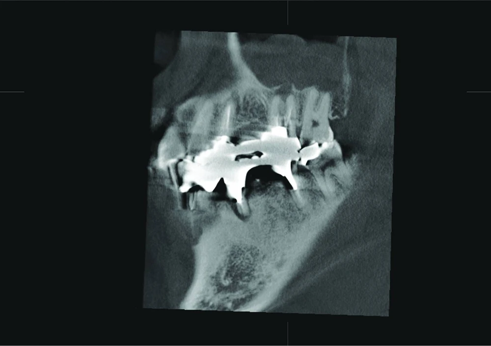
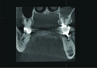
The sagittal view clearly confirms perforation of the Schneiderian membrane, and the coronal view revealed an odontogenic maxillary sinusitis and pachymenia. The injury of the sinus membrane may have been overlooked in this case if the diagnosis had been formulated on the basis of an X-ray that did not show the problem so clearly.
Bài đăng lần đầu ngày: 18 Tháng 5, 2018 @ 7:15 chiều