Xác định chiều dài làm việc: sai lầm và hậu quả
The Apical Terminus: A Comprehensive Clinical Review of Working Length Determination, Procedural Errors, and Treatment Outcomes in Endodontics
Section 1: The Clinical Imperative of Working Length
The success of endodontic therapy is predicated on the thorough chemo-mechanical debridement and three-dimensional obturation of the root canal system. Central to achieving this objective is the accurate determination of the working length (WL). Formally defined as "the distance from a coronal reference point to the point at which canal preparation and obturation should terminate," the working length represents the longitudinal extent of treatment and is unequivocally one of the most crucial factors in determining the prognosis of root canal therapy.1 Improper measurement of this critical dimension is a primary contributor to both short-term post-operative complications and long-term treatment failure, making its precise establishment a foundational pillar of modern endodontic practice.5 The clinical significance of working length is rooted in a fundamental biological principle: the confinement of all procedures within the intracanal space. The root canal system can be conceptualized as a "biological container," with the cementodentinal junction (CDJ) representing the histological transition from the internal, pulpal environment (endodontium) to the external, periradicular tissues (periodontium).6 The primary goal of accurate WL determination is to respect this biological boundary. Errors in this measurement are not merely geometric inaccuracies; they are biological violations that either leave a pathogenic reservoir within the canal (when working short) or induce iatrogenic trauma to the periapical tissues (when working long). Therefore, the act of measuring WL is an attempt to define and honor the host's biological integrity, a failure of which directly precipitates inflammation, persistent infection, and the ultimate failure of therapy. The accuracy of the working length has a direct and profound impact on the efficacy of each subsequent phase of treatment. During chemo-mechanical debridement, a correct WL ensures that shaping instruments can access and clean the entire length of the infected canal system, particularly the critical apical third, without inadvertently transporting pathogens and debris into the periapical space.3 This, in turn, facilitates effective disinfection by allowing irrigants, such as Sodium hypochlorite, to penetrate and act upon microorganisms harbored in the complex anatomical ramifications of the apical delta.9 Finally, for obturation, a precisely determined apical terminus is a prerequisite for creating a dense, hermetic seal. It allows for the controlled compaction of filling materials to create a three-dimensional fluid-tight barrier against microleakage and reinfection, preventing the extrusion of materials that can act as foreign body irritants to the periapical tissues.6 The correlation between working length accuracy and long-term success is well-documented. Endodontic failures are frequently attributed to errors in WL, which lead to under-extended or over-extended root fillings.5 According to established guidelines, filling the root canal more than 2 mm short of the radiographic apex or extending beyond it is considered a technical failure that severely compromises the prognosis.3 This concept is encapsulated in the "pyramid of success" in endodontics, which posits that small errors committed during the initial stages of treatment, such as WL determination, can cascade and have a disproportionately negative influence on the final outcome.12
Section 2: Anatomy of the Apical Third: The Clinician's Roadmap
Navigating the apical third of the root canal system requires a comprehensive understanding of its complex and highly variable anatomy. This region, often referred to as the apical "significant zone," is not a simple terminus but a complex of distinct anatomical and histological landmarks that rarely coincide, presenting a formidable challenge to the clinician.7 The core difficulty in working length determination stems from what can be termed an "Anatomical Uncertainty Principle": the ideal biological target for instrumentation, the apical constriction, is a histological landmark that is invisible on radiographs and cannot be detected with clinical certainty. Clinicians are therefore compelled to use imperfect proxies—radiographic images and electronic impedance measurements—to estimate the location of this unseen destination. This inherent uncertainty is the fundamental reason why multiple determination methods are advocated and why procedural errors remain a persistent clinical challenge.
Deconstructing the Apical Complex: Foramen, Constriction, and Junctions
The apical complex comprises several key landmarks, each with a distinct definition and clinical relevance 2:
- Anatomic Apex: The morphological tip or end of the root as determined by its physical shape.
- Radiographic Apex: The tip of the root as visualized on a two-dimensional radiograph. Due to image distortion and root morphology, its location can vary significantly from the true anatomic apex.6
- Apical Foramen (Major Diameter): The main apical opening of the root canal on the external root surface. It is frequently located eccentric to the anatomic apex, with studies reporting deviations in up to 80% of teeth.7 The mean distance between the major foramen and the anatomic apex is approximately 0.59 mm.7
- Apical Constriction (Minor Diameter): The narrowest portion of the root canal, located coronally to the apical foramen. Its position is variable but is generally considered to be 0.5 mm to 1.5 mm short of the major foramen.2
- Cementodentinal Junction (CDJ): The histological point where the dentin of the canal wall meets the cementum of the external root surface. Its position is highly variable, ranging from 0.5 mm to 3.0 mm from the anatomic apex.6 It is critical to recognize that the CDJ and the apical constriction are not anatomically synonymous, though they may be located near one another.7
The Apical Constriction as the Ideal Physiological Terminus
There is a broad consensus within the endodontic community that the apical constriction should be the target for the termination of canal preparation and obturation.7 The rationale for selecting this landmark as the ideal physiological terminus is threefold. First, it generally represents the transition from pulpal tissue to periodontal tissue. Second, as the narrowest diameter of the canal, it provides a natural stop or abutment against which obturation materials can be condensed, minimizing the risk of extrusion. Third, terminating procedures at the constriction creates the smallest possible wound site at the interface with the periradicular tissues, which is the most favorable condition for predictable healing.6
Anatomical Variability and Its Clinical Implications
The primary challenge in locating the apical constriction is its profound anatomical variability. The distance between the constriction and the anatomic apex can range from 0 mm to as much as 4 mm.14 This relationship is not static and is influenced by a host of physiological and pathological factors. Age-related deposition of cementum can shift the position of the foramen over time. Pathological processes such as inflammatory root resorption can partially or completely destroy the natural apical constriction, leaving a wide, open apex that is difficult to manage.15 Furthermore, the apical third frequently contains complex morphological features, including accessory canals, bifurcations, and apical deltas, which can harbor microorganisms. If instrumentation falls short of the full canal length, these areas remain untreated and can serve as a nidus for persistent infection.16 This anatomical unpredictability renders any determination method based on "average values" clinically unacceptable and underscores the need for a patient-specific approach.14
Reconciling Radiographic, Anatomic, and Electronic Apices
To navigate this complex anatomy, the clinician must reconcile information from different sources. The "radiographic apex" is a two-dimensional shadow, the "anatomic apex" is a physical structure, and the "electronic apex" is a location determined by electrical impedance, which typically corresponds to the apical foramen.1 The clinical objective is to synthesize the data from radiographic and electronic methods to infer the position of the unseen histological landmark—the apical constriction—and thereby establish a biologically sound working length.
Section 3: A Comparative Analysis of Working Length Determination Methodologies
The evolution of endodontic practice has seen a shift from reliance on a single, often fallible, method of working length determination to a multi-technique approach that leverages the strengths of different technologies. The modern standard of care is not about selecting the single "best" method but about employing a strategy of "Synergistic Triangulation." Each technique—radiographic, electronic, and tomographic—possesses inherent flaws and limitations. However, when used in combination, they compensate for one another's weaknesses, allowing the clinician to triangulate the position of the apical terminus with a degree of accuracy and confidence that no single method can provide alone. This strategy involves using radiographs for anatomical planning, electronic apex locators for precise apical navigation, and advanced imaging for complex cases, creating a layered, data-rich approach to a fundamentally challenging clinical task.
The Radiographic Standard: Techniques, Advantages, and Inherent Limitations
For decades, radiography has been the cornerstone of working length determination. The most common technique involves placing an endodontic file into the canal to an estimated length and taking a periapical radiograph, often referred to as an "instrument contrast radiograph".14
- Techniques: The paralleling technique, where the film or sensor is placed parallel to the long axis of the tooth, is generally considered more accurate and produces less geometric distortion than the bisecting-angle technique.17 Various methods, such as that described by Ingle, use the radiographic apex as a reference, with the working length typically established 0.5 mm to 1.0 mm short of this point to approximate the location of the apical constriction.5
- Advantages: Despite its limitations for precise length measurement, radiography provides invaluable diagnostic information that is unobtainable by other means. It reveals root and canal morphology, the presence and severity of curvatures, canal calcifications, the number of roots, and the status of the periapical tissues.5
- Limitations: The fundamental flaw of radiography is its two-dimensional representation of a three-dimensional object. This leads to the superimposition of anatomical structures, making it difficult to visualize canals or foramina located on the buccal or lingual aspects of the root.1 Studies have shown that even with a radiographically acceptable file position, the instrument tip may be extended beyond the apical foramen in up to 28.5% of cases.17 Additional limitations include image distortion, variability in interpretation among clinicians, and patient exposure to ionizing radiation.5
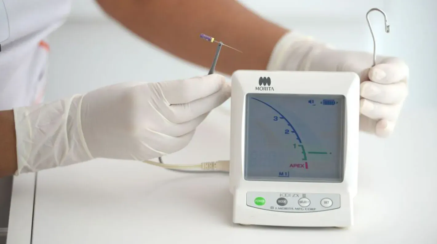
The Electronic Revolution: Principles and Evolution of Electronic Apex Locators (EALs)
The introduction and refinement of electronic apex locators (EALs) have revolutionized working length determination, offering a dynamic and highly accurate method for locating the apical terminus.
- Principles and Evolution: EALs operate on the principle that the human body completes an electrical circuit, with the periodontal ligament and oral mucous membrane having a relatively constant electrical resistance of approximately $6.5 kOmega$.20 Early-generation devices were resistance-based and operated on direct current, making them highly susceptible to inaccuracies in the presence of conductive fluids (e.g., irrigants, blood, pus) within the canal.21 Modern EALs are impedance-based, using multiple alternating current frequencies to measure and compare the electrical impedance as the file tip approaches the apical foramen. This technology is significantly less affected by the canal contents, providing reliable readings in both wet and dry environments.1 The most recent "adaptive" EALs can continuously measure canal humidity and adjust their algorithm accordingly, further enhancing precision.21
- Accuracy and Advantages: Numerous in vivo and in vitro studies have demonstrated the high accuracy of modern EALs, which can locate the apical foramen with greater than 90% accuracy.1 Multiple comparative studies have concluded that EALs are more accurate than radiography alone for determining working length.4 The primary advantages of EALs are their high degree of precision, the significant reduction in the number of intra-operative radiographs required (thereby reducing patient radiation exposure), and increased clinical efficiency.13
Advanced Imaging: The Role of Cone-Beam Computed Tomography (CBCT)
Cone-beam computed tomography (CBCT) provides a three-dimensional visualization of the dentoalveolar complex, overcoming the inherent limitations of 2D radiography.9 While not routinely used for WL determination in uncomplicated cases due to its higher radiation dose and cost, CBCT is an invaluable tool for complex situations, such as teeth with unusual anatomy, suspected perforations, or extensive resorptive defects. Studies have shown that WL measurements obtained from CBCT images are highly precise and are not statistically different from those obtained with EALs.27
Ancillary and Adjunctive Techniques: Tactile Sensation and Paper Point Evaluation
While largely superseded by electronic methods, some traditional techniques remain useful as adjunctive aids.
- Tactile Sensation: This involves the clinician's ability to feel the file tip passing through the apical constriction. While a valuable skill, it is highly operator-dependent and is considered unreliable in canals that are curved, sclerotic, or have immature apices.1
- Paper Point Evaluation: This technique is based on observing moisture or blood on the tip of a paper point inserted to the estimated WL. A dry point indicates the length is short, while a point wet with blood or exudate suggests the length is long. This can be a useful confirmatory check, particularly before obturation.1
Table 1: Comparative Analysis of Working Length Determination Methods
Parameter Conventional/Digital Radiography Electronic Apex Locator (EAL) Cone-Beam Computed Tomography (CBCT) Principle of Operation 2D X-ray imaging of file position relative to radiographic apex. Measurement of electrical impedance between a file in the canal and an oral mucosal electrode. 3D volumetric X-ray imaging of tooth and surrounding structures. Reported Accuracy 72-96% within $pm 0.5$ mm of actual length. Accuracy is technique-dependent.4 >90-98% within $pm 0.5$ mm of the apical foramen. Generally considered more accurate than radiography.1 High precision, comparable to EALs. Not statistically superior in all studies.27 Radiation Exposure Low to moderate per image. Cumulative dose can be a concern with multiple exposures.19 None. Significantly reduces the need for intra-operative radiographs.20 Significantly higher than periapical radiography. Use should be justified.27 Influence of Canal Contents Not directly affected, but fluid-filled canals may have less contrast. Early generations were inaccurate in presence of electrolytes. Modern impedance-based EALs are highly reliable in wet canals.21 Not affected. Provides clear visualization regardless of canal contents. Key Limitations 2D image of 3D structure, geometric distortion, anatomical superimposition, observer variability.13 Provides no anatomical context (e.g., curvature). Can be inaccurate with open apices, perforations, or contact with metallic restorations.1 Higher radiation dose, cost, potential for artifacts from metallic objects. Not a routine tool for WL.27 Primary Clinical Role Initial diagnosis, anatomical assessment (macro-map), and confirmation of final obturation. Precise, real-time navigation to the apical foramen (micro-map) during instrumentation. Diagnosis and treatment planning for complex cases with anatomical anomalies, trauma, or resorption.
Section 4: The Perils of Inaccuracy: Under-instrumentation and Its Sequelae
Establishing a working length that is short of the true canal terminus is a critical procedural error that initiates a predictable cascade of iatrogenic events, culminating in treatment failure. This sequence, which can be described as the "Apical Domino Effect," begins with an initial measurement error and progresses through canal blockage and ledge formation to the ultimate retention of a pathogenic reservoir. Each step in this cascade directly and predictably leads to the next, transforming a simple inaccuracy into a complex and often insurmountable clinical problem.
"Working Short": The Pathophysiology of Retained Pathogens
The immediate and most significant biological consequence of under-instrumentation is the incomplete debridement of the apical portion of the root canal system.1 When instrumentation and irrigation fall short, necrotic tissue, microorganisms, and their toxic byproducts are left undisturbed in the most anatomically complex region of the canal.5 This untreated apical segment, often containing deltas and accessory canals, becomes a protected reservoir for persistent infection. The host's immune defenses cannot clear pathogens from within this sealed-off space, leading to the establishment or continuation of periradicular inflammation and pathology.12 Studies have demonstrated that even a 1 mm loss in working length can increase the probability of endodontic failure by 14% in teeth with pre-existing apical periodontitis.5
Iatrogenic Blockages: The Creation and Management of Dentin Plugs
The mechanical action of endodontic files within the canal produces dentin chips and grinds pulpal remnants. When working short, these debris, referred to as "dentin mud," are not flushed from the canal but are instead transported apically and compacted against the un-instrumented portion of the canal wall.16 Progressively larger instruments further condense this debris, forming a dense plug that effectively blocks the apical terminus.12 Once a dentin plug is formed, it becomes extremely difficult for subsequent instruments or irrigants to penetrate to the full working length. This loss of working length is a common procedural complication that effectively seals pathogens within the most critical part of the canal system, dooming the treatment to failure.12
Ledge Formation: Etiology and Strategies for Bypass
Faced with a blocked canal and a loss of working length, a common clinical error is to apply excessive apical force with a relatively stiff instrument in an attempt to regain the original canal path. This action rarely succeeds in dislodging the plug; instead, the instrument is deflected by the blockage and begins to carve a new, false path into the dentin wall, creating a "ledge".16 Incorrect working length determination is cited as a primary cause of ledge formation.31 A ledge is a significant iatrogenic error that creates an artificial barrier, further preventing access to the apical portion of the canal. Navigating past a ledge is a technically demanding procedure that requires the use of small, pre-curved files and a delicate touch; often, the original canal path cannot be renegotiated, making complete debridement impossible.31
The Untreated Apex: A Reservoir for Persistent Infection and Treatment Failure
The culmination of the "Apical Domino Effect"—an initially short working length leading to blockage and subsequent ledge formation—is an incompletely cleaned, shaped, and sealed root canal system. This is a principal cause of endodontic failure. The untreated apical segment remains a persistent source of microbial irritation to the periradicular tissues, resulting in post-operative symptoms, the non-healing of existing periapical lesions, or the development of new pathology.5 The final radiograph may appear to show a well-filled canal, but the biological reality is a failure to achieve the primary objective of endodontic therapy: the elimination of infection from the entire root canal system.
Section 5: The Perils of Inaccuracy: Over-instrumentation and Periapical Trauma
While working short leads to failure through retained infection, establishing a working length that extends beyond the apical constriction results in failure through iatrogenic trauma and the violation of the periradicular tissues. This error breaches the biological boundary of the tooth, introducing mechanical, chemical, and microbial insults directly into the periodontal ligament and alveolar bone. The consequences range from acute post-operative pain to permanent anatomical damage and compromised long-term healing.
"Working Long": Breaching the Periradicular Tissues
Over-instrumentation is the extension of endodontic files and irrigants beyond the apical foramen.1 This action mechanically injures the delicate periapical tissues, causing hemorrhage into the canal and triggering an acute inflammatory response characterized by the release of chemical mediators.22 This trauma is a common cause of significant post-operative pain and discomfort.19 Furthermore, instrumentation beyond the apex can inadvertently transport intracanal bacteria and debris into the sterile periapical region, seeding a new infection or exacerbating an existing one, which can delay or prevent healing.3
Apical Transportation and Perforation: Altering Anatomy and Compromising the Seal
Repeated instrumentation beyond the apex, particularly with larger and less flexible stainless steel files in curved canals, can permanently alter the apical anatomy. The inherent tendency of these instruments to straighten themselves results in the preferential removal of dentin from the outer wall of the canal's curvature. This leads to the transportation of the apical foramen, creating an elliptical or "teardrop" shaped opening, a phenomenon often called "zipping".12 This iatrogenic enlargement destroys the natural apical constriction, which is the narrowest point of the canal and the key to achieving a controlled, three-dimensional obturation. Without this "apical stop," it becomes exceedingly difficult to contain filling materials within the canal, compromising the apical seal.32 In severe cases, aggressive over-instrumentation can lead to a frank perforation of the root, creating a direct communication between the canal system and the periodontium.31
Overfilling vs. Overextension: A Critical Distinction for Prognosis
A crucial nuance exists in the prognosis of cases where obturation material is extruded beyond the apex, a phenomenon that can be termed the "Overfilling Paradox." The radiographic presence of extruded material is, by itself, a poor predictor of the clinical outcome. Success is determined not by the absence of extrusion but by the quality of the apical seal.
- Overfilling is defined as the extrusion of a small amount of sealer or gutta-percha beyond the apex when the canal itself has been thoroughly cleaned, shaped, and is hermetically sealed at the apical terminus.11
- Overextension describes the extrusion of material when the canal is inadequately cleaned or sealed, leaving voids that permit bacterial leakage from the canal system into the periapex.11
While overextension is strongly correlated with treatment failure, numerous long-term case series have shown that cases of true overfilling can heal successfully. The periapical tissues appear to have a remarkable capacity to either resorb or encapsulate small amounts of sterile filling material, provided that the source of infection—the root canal—is completely sealed.11 This demonstrates that the failure is not caused by the material outside the tooth, but by the persistent bacteria left inside due to an inadequate seal.
Clinical Consequences: Post-Operative Pain, Foreign Body Reactions, and Nerve Injury
Extruded gutta-percha and, more significantly, root canal sealers can act as foreign bodies, inciting a chronic inflammatory response or a foreign body giant cell reaction in the periapical tissues, which may mimic endodontic failure and require surgical removal.11 A particularly severe complication of overfilling, especially in mandibular premolars and molars, is the extrusion of material into the mandibular canal. This can cause direct mechanical compression or chemical neurotoxicity to the inferior alveolar nerve, resulting in debilitating and sometimes permanent neurological deficits such as paresthesia (tingling or numbness) of the lip, chin, and gingiva.38 Similarly, extrusion of material from maxillary posterior teeth into the maxillary sinus can lead to foreign body reactions and chronic sinusitis.38 Table 2: Summary of Adverse Events Associated with Incorrect Working Length
Parameter Consequences of "Working Short" (Under-instrumentation) Consequences of "Working Long" (Over-instrumentation) Primary Biological Error Incomplete debridement and disinfection; retention of necrotic and infected tissue within the canal.1 Mechanical, chemical, and microbial injury to periradicular tissues; violation of the biological apex.1 Key Iatrogenic Mishaps Apical blockage with dentin plug, loss of working length, ledge formation, instrument separation.12 Apical transportation ("zipping"), apical perforation, extrusion of irrigants and obturation materials.12 Impact on Apical Anatomy Original canal anatomy is blocked and un-instrumented; a false canal path may be created by a ledge.16 The natural apical constriction is destroyed; the foramen is widened and transported from its original position.32 Primary Clinical Sequelae Persistent intracanal infection, failure of periapical lesion to heal, development of new periapical pathology.5 Post-operative pain and inflammation, foreign body reaction, potential for nerve damage (paresthesia), sinus perforation.22 Post-Operative Symptoms Often delayed or persistent low-grade discomfort; may be asymptomatic until a flare-up occurs.19 Often characterized by acute and severe post-operative pain, swelling, and tenderness to percussion.19 Long-Term Prognosis Poor. High likelihood of endodontic failure due to persistent microbial infection.5 Variable. Depends on the extent of damage and quality of the apical seal. Overextension is linked to failure; true overfilling may heal successfully.11
Section 6: Management and Mitigation of Working Length Errors
The successful management of iatrogenic errors stemming from incorrect working length determination requires prompt recognition, a thorough understanding of the underlying problem, and the application of specific clinical protocols. In cases of severe anatomical alteration, such as the destruction of the apical constriction, management represents a paradigm shift from conventional obturation to a process of "Apical Reconstruction." This approach involves not merely filling a pre-existing space but actively engineering a new biological or artificial barrier at the root apex using advanced bioactive materials, thereby transforming a compromised tooth into a salvageable one.
Clinical Protocols for Correcting Procedural Mishaps
The approach to managing a procedural error depends on its nature and severity.
- Bypassing Ledges: A ledge created by working short and applying excessive force can sometimes be bypassed. This is typically attempted by using a small (e.g., #08 or #10 K-file) with a distinct curve placed at its apical tip. The file is carefully "hunted" along the canal wall opposite the ledge to find the entrance to the original canal path. Copious irrigation and lubrication are essential. Once the original canal is located and patency is re-established, it is carefully instrumented to create a smooth glide path before proceeding with larger shaping files.10
- Managing Minor Over-instrumentation: If instrumentation with small files inadvertently extends beyond the apex, it often precipitates bleeding into the canal. In many cases, this can be managed by completing the instrumentation at the corrected working length while using copious irrigation with sodium hypochlorite, which aids in hemostasis. A short-term intracanal dressing of calcium hydroxide may also be recommended to reduce inflammation before obturation.32
- Retreatment of Overfilled Cases: If an overfilled tooth becomes symptomatic or shows signs of a non-healing lesion, retreatment may be indicated. Non-surgical approaches involve removing the existing gutta-percha and attempting to retrieve the extruded segment using specialized instruments like H-files or ultrasonic tips, often under the magnification of a dental operating microscope.42 If non-surgical retrieval is not possible or fails to resolve symptoms, surgical intervention (apicoectomy) to remove the extruded material and seal the root-end may be necessary. In select cases, intentional replantation has also been used as a treatment modality.36
Management of the Iatrogenically Open Apex: Apical Barrier Techniques
When significant over-instrumentation destroys the apical constriction and creates a wide, parallel, or flared apex, conventional obturation techniques are ineffective, as there is no stop to contain the filling materials.33 The modern management for these cases involves creating an artificial apical barrier, or "plug," against which the remainder of the canal can be filled.
- Bioceramic Materials: The materials of choice for this procedure are bioactive bioceramics, primarily Mineral Trioxide Aggregate (MTA) and Biodentine™. These materials are biocompatible, have excellent sealing ability, and are capable of inducing hard tissue formation. A 3 mm to 5 mm plug of MTA or a similar material is carefully placed and compacted into the apical extent of the canal to create a durable artificial barrier.33 The placement of an internal matrix, such as a collagen membrane or platelet-rich fibrin (PRF), just beyond the apex can help prevent extrusion of the bioceramic during placement.48
- Reinforcement of Weakened Roots: Aggressive over-instrumentation can not only widen the apex but also excessively flare the coronal portion of the canal, thinning the radicular dentin walls and making the tooth susceptible to vertical root fracture.33 In such cases, after placement of the apical plug, the weakened root structure can be internally reinforced. This involves bonding a thick layer of adhesive material, such as a composite resin or giomer, into the canal space, often around a fiber post, to strengthen the tooth before the final coronal restoration is placed.33
Long-Term Follow-Up and Prognostic Assessment of Compromised Cases
Any tooth that has experienced a significant procedural error related to working length requires diligent long-term follow-up, including periodic clinical and radiographic examinations.11 The prognosis is variable and depends heavily on the nature and extent of the initial error, the success of the corrective procedure, the pre-operative pulpal and periapical diagnosis, and the quality of the final coronal seal. While modern techniques have made it possible to salvage many of these compromised teeth, the prognosis is generally considered to be lower than for uncomplicated cases.51
Section 7: Best Practices and Future Horizons in Working Length Determination
Achieving a consistently accurate and predictable working length is the result of a systematic clinical protocol that integrates multiple technologies and acknowledges their respective limitations. The evolution of this process is now moving beyond simple measurement toward a "Data-Driven Apex" approach. This emerging paradigm de-emphasizes reliance on a single reading or subjective feel, focusing instead on creating a continuous, multi-source feedback loop of data points gathered throughout the procedure. By triangulating information from radiographs, electronic locators, tactile feedback, and, in the future, artificial intelligence, clinicians can build a more robust and dynamic model of the apical anatomy for each canal, providing the most effective strategy to overcome the fundamental "Anatomical Uncertainty Principle."
A Protocol for Achieving Predictable and Accurate Working Length
A best-practice protocol for working length determination involves a synergistic sequence of steps: 1. Pre-operative Radiographic Analysis: A high-quality, pre-operative parallel radiograph is essential for initial diagnosis and treatment planning. It provides the "macro-map" of the tooth, revealing the number of roots, estimated canal length, degree of curvature, and presence of any calcifications or anatomical anomalies.1 2. Straight-Line Access and Coronal Pre-flaring: The access cavity must be designed to provide unobstructed, straight-line access to each canal orifice. Following this, the coronal two-thirds of the canal should be "pre-flared" with orifice shapers or rotary files. This step removes coronal interferences, allowing subsequent instruments to pass more easily and passively into the apical third, which enhances tactile sensation and the accuracy of EAL readings.1 3. Electronic Length Determination with a Patency File: With the canal filled with an irrigant, a small, flexible K-file (e.g., #10 or #15) is used to negotiate the canal to its terminus while attached to an EAL. This establishes the "electronic length" to the major foramen. The working length is then typically set 0.5 mm to 1.0 mm short of this measurement to approximate the apical constriction.1 Maintaining apical patency with a small file throughout the procedure helps prevent blockage.16 4. Constant Verification: The working length is not a static measurement. In curved canals, instrumentation can cause straightening, which effectively shortens the canal length. Therefore, the WL should be periodically re-verified with the EAL throughout the shaping procedure.1 5. Radiographic Confirmation: A final working length confirmation radiograph with the master apical file or master obturation cone in place is recommended to verify the length and its relationship to the overall root anatomy before final obturation.29
Troubleshooting Common Issues with Electronic Apex Locators
Clinicians should be aware of common situations that can lead to inaccurate EAL readings and know how to manage them 1:
- Erratic or "Instant" Readings: Often caused by an electrical short circuit. This can occur if the pulp chamber is flooded with irrigant, if the file contacts a metallic restoration (crown, amalgam), or if there is gingival tissue contact. Management involves drying the chamber, isolating the file from the metal with a plastic instrument or composite, and ensuring good rubber dam isolation.
- Slow or Delayed Readings: May indicate a blocked or heavily calcified canal that is impeding the electrical path. Management involves recapitulating with a small file to clear debris or using a chelating agent to help negotiate the calcification. An extremely dry canal can also cause a slow reading; the canal should be moistened, preferably with saline.
- Inaccurate Reading in Wide Apices: In cases with an immature or resorbed apex, a small file may not make adequate contact with the canal walls. Using a larger file that more closely approximates the apical diameter will provide a more stable and accurate reading.13
The Future Landscape: Artificial Intelligence and Enhanced Imaging
The next frontier in working length determination lies in the integration of advanced computational analysis and enhanced imaging.
- Artificial Intelligence (AI): The application of machine learning, particularly deep learning models like convolutional neural networks (CNNs), is showing remarkable potential in endodontics. AI algorithms are being trained to analyze 2D radiographs and 3D CBCT scans to automatically detect the apical foramen, measure canal length, identify root fractures, and trace complex canal morphologies.52 By automating these tasks and providing an objective analysis, AI has the potential to reduce operator-dependent errors, improve diagnostic accuracy, and enhance the predictability of treatment planning.
- Enhanced Imaging Tools: The continued development of software for digital radiography and CBCT will provide clinicians with more powerful visualization tools. Image processing modules that can enhance contrast, apply filters, or render images in color can improve the visibility of fine anatomical details, further aiding in the interpretation of the complex apical region.27
Together, these advancements promise to provide clinicians with an even more robust dataset, further refining the "Data-Driven Apex" approach and moving the practice of endodontics ever closer to the goal of predictable, error-free treatment. Nguồn trích dẫn 1. The working length – Style Italiano Endodontics, truy cập vào tháng 10 26, 2025, https://endodontics.styleitaliano.org/the-working-length/ 2. working length | PPTX – Slideshare, truy cập vào tháng 10 26, 2025, https://www.slideshare.net/slideshow/working-length-70030221/70030221 3. Comparative Evaluation of Root Canal Working Length …, truy cập vào tháng 10 26, 2025, https://jdmt.mums.ac.ir/article_17970_0be14a8471619d8b8a8329f30aa8c04b.pdf 4. (PDF) Comparative evaluation of the accuracy of working length …, truy cập vào tháng 10 26, 2025, https://www.researchgate.net/publication/372983727_Comparative_evaluation_of_the_accuracy_of_working_length_determination_by_radiographic_method_and_electronic_apex_locator 5. (PDF) significance of working length in rct – ResearchGate, truy cập vào tháng 10 26, 2025, https://www.researchgate.net/publication/375799867_significance_of_working_length_in_rct 6. 14 Electronic Apex Locators and Conventional Radiograph in …, truy cập vào tháng 10 26, 2025, https://pocketdentistry.com/14-electronic-apex-locators-and-conventional-radiograph-in-working-length-measurement/ 7. Apical reference point – A clinical perspective – Journal of Academy …, truy cập vào tháng 10 26, 2025, https://adejournal.com/apical-reference-point-a-clinical-perspective/ 8. The Endodontics Root Canals Instrumentation & Irrigation & Obturation, truy cập vào tháng 10 26, 2025, https://www.uobabylon.edu.iq/eprints/publication_4_2811_1726.pdf 9. Root Canal Instrumentation: Current Trends and Future Implications, truy cập vào tháng 10 26, 2025, https://jicrcr.com/index.php/jicrcr/article/download/748/543/1564 10. An Update on Root Canal Preparation Techniques and How to Avoid Procedural Errors in Endodontics – The Open Dentistry Journal, truy cập vào tháng 10 26, 2025, https://opendentistryjournal.com/VOLUME/15/PAGE/318/ 11. The fate of overfilling in root canal treatments with long-term follow-up: a case series – PMC, truy cập vào tháng 10 26, 2025, https://pmc.ncbi.nlm.nih.gov/articles/PMC8170384/ 12. Procedural errors in endodontic treatment: analysis of preclinical …, truy cập vào tháng 10 26, 2025, https://www.termedia.pl/Procedural-errors-in-endodontic-treatment-analysis-of-preclinical-step-back-preparation-performed-on-training-blocks,137,40152,1,1.html 13. Evaluation of the Accuracy of Electronic Apex Locators in Modern …, truy cập vào tháng 10 26, 2025, https://www.mdpi.com/1648-9144/60/10/1709 14. 19 Determination of Working Length | Pocket Dentistry, truy cập vào tháng 10 26, 2025, https://pocketdentistry.com/19-determination-of-working-length/ 15. 9: Problem Solving in Working Length Determination | Pocket Dentistry, truy cập vào tháng 10 26, 2025, https://pocketdentistry.com/9-problem-solving-in-working-length-determination/ 16. Working Length: The Pitfalls of Working Short & The Box Preparation, truy cập vào tháng 10 26, 2025, https://www.endoruddle.com/blogs/show/10/working-length-the-pitfalls-of-working-short-the-box-preparation 17. (PDF) Radiographic assessment of endodontic working length, truy cập vào tháng 10 26, 2025, https://www.researchgate.net/publication/276021399_Radiographic_assessment_of_endodontic_working_length 18. A comparative evaluation of electronic and radiographic determination of root canal length in primary teeth: An in vitro study – NIH, truy cập vào tháng 10 26, 2025, https://pmc.ncbi.nlm.nih.gov/articles/PMC3636831/ 19. Endodontic tips for working length and apex locator accuracy, truy cập vào tháng 10 26, 2025, https://www.ildentistamoderno.com/endodontic-tips-for-working-length-and-apex-locator-accuracy/ 20. Apex Locators in Root Canal Treatment – Endodontic Partners, truy cập vào tháng 10 26, 2025, https://sereneendo.com/endodontic-technology/apex-locators/ 21. Electronic apex locator – Wikipedia, truy cập vào tháng 10 26, 2025, https://en.wikipedia.org/wiki/Electronic_apex_locator 22. Effect of Working Length Measurement by Electronic Apex Locater …, truy cập vào tháng 10 26, 2025, https://pjmhsonline.com/2021/feb/736.pdf 23. Reliability of different methods of measurement … – MedCrave online, truy cập vào tháng 10 26, 2025, https://medcraveonline.com/JDHODT/JDHODT-15-00617.pdf 24. Working Length Determination of Root Canal of Young Permanent Tooth: An In vitro Study – PMC – NIH, truy cập vào tháng 10 26, 2025, https://pmc.ncbi.nlm.nih.gov/articles/PMC4160679/ 25. An in vitro Comparison of Root Canal Measurement in Permanent …, truy cập vào tháng 10 26, 2025, https://wjoud.com/doi/WJOUD/pdf/10.5005/jp-journals-10015-1104 26. A comparative evaluation of electronic and radiographic … – NIH, truy cập vào tháng 10 26, 2025, https://www.ncbi.nlm.nih.gov/pmc/articles/PMC3636831/ 27. Advances in Working Length Determination – ResearchGate, truy cập vào tháng 10 26, 2025, https://www.researchgate.net/publication/364069677_Advances_in_Working_Length_Determination 28. Working Length Determination Using Cone-Beam Computed …, truy cập vào tháng 10 26, 2025, https://pubmed.ncbi.nlm.nih.gov/27471524/ 29. Mastering Working Length Determination in Endodontics – YouTube, truy cập vào tháng 10 26, 2025, https://www.youtube.com/watch?v=fQtf5xzqU9I 30. Tips for successfuL LengTh DeTerminaTion – Dentsply Sirona, truy cập vào tháng 10 26, 2025, https://www.dentsplysirona.com/content/dam/master/regions-countries/north-america/product-procedure-brand/endodontics/product-categories/endodontic-equipment/apex-locators/promark-al/documents/END-TipCard-ProMark-AL-EN.pdf.pdf 31. Errors in root canal preparation: a review of the literature and clinical …, truy cập vào tháng 10 26, 2025, https://scielo.org.za/scielo.php?script=sci_arttext&pid=S0011-85162019000500008 32. Overinstrumentation: Treatment, truy cập vào tháng 10 26, 2025, https://secure.dentistry.ubc.ca/intranet/visuendo-svn/m9/overinstrumentation-treatment.html 33. Management of a Flared Root Canal with an Iatrogenically Widened Apex – SciSpace, truy cập vào tháng 10 26, 2025, https://scispace.com/pdf/management-of-a-flared-root-canal-with-an-iatrogenically-37cmp1hf75.pdf 34. (PDF) The fate of overfilling in root canal treatments with long-term follow-up: a case series, truy cập vào tháng 10 26, 2025, https://www.researchgate.net/publication/351329055_The_fate_of_overfilling_in_root_canal_treatments_with_long-term_follow-up_a_case_series 35. The fate of overfilling in root canal treatments with long-term follow-up: a case series, truy cập vào tháng 10 26, 2025, https://www.semanticscholar.org/paper/The-fate-of-overfilling-in-root-canal-treatments-a-Malagnino-Pappalardo/2df71a91b4326c0a745566551a97e5c445b51d62 36. Surgical Resolution of Chronic Tissue Irritation Caused by Extruded Endodontic Filling Material – Canadian Dental Association, truy cập vào tháng 10 26, 2025, https://www.cda-adc.ca/jcda/vol-71/issue-7/487.pdf 37. Extrusion of Endodontic Filling Materials: Medico-Legal Aspects. Two Cases – PMC – NIH, truy cập vào tháng 10 26, 2025, https://pmc.ncbi.nlm.nih.gov/articles/PMC2697057/ 38. Root Canal Overfill Symptoms and Complications – Spetsas Buist, truy cập vào tháng 10 26, 2025, https://www.spetsasbuist.com/blog/root-canal-overfill-symptoms/ 39. The Prognosis of Altered Sensation after Extrusion of Root Canal Filling Materials: A Systematic Review of the Literature | Request PDF – ResearchGate, truy cập vào tháng 10 26, 2025, https://www.researchgate.net/publication/301758748_The_Prognosis_of_Altered_Sensation_after_Extrusion_of_Root_Canal_Filling_Materials_A_Systematic_Review_of_the_Literature 40. Management of Gutta-Percha over Filling of Mandibular First Molar By Intentional Replantation: Case Report, truy cập vào tháng 10 26, 2025, https://www.ijmsdr.org/published%20paper/li1i15/Management-of-Gutta-Percha-over-Filling-of-Mandibular-First-Molar-By-Intentional-Replantation-Case-Report.pdf 41. Management of iatrogenic errors of root canal treatment- A case with multiple complex mishaps, ledge and perforation management – Style Italiano Endodontics, truy cập vào tháng 10 26, 2025, https://endodontics.styleitaliano.org/management-of-iatrogenic-errors-of-root-canal-treatment-a-case-with-multiple-complex-mishaps-ledge-and-perforation-management/ 42. A Nonsurgical Approach for Removal of Overfilling Guttapercha: Case Report, truy cập vào tháng 10 26, 2025, https://pubs.sciepub.com/ijdsr/9/1/5/index.html 43. Overfill Resolution Through Non-Surgical Endodontic Treatment: A Case Report – PMC, truy cập vào tháng 10 26, 2025, https://pmc.ncbi.nlm.nih.gov/articles/PMC12318327/ 44. Endodontic management of a tooth with apical overfilling and perforating external root resorption: A case report – PMC – NIH, truy cập vào tháng 10 26, 2025, https://pmc.ncbi.nlm.nih.gov/articles/PMC7752420/ 45. Management of a Flared Root Canal with an Iatrogenically Widened Apex – SciSpace, truy cập vào tháng 10 26, 2025, https://scispace.com/papers/management-of-a-flared-root-canal-with-an-iatrogenically-37cmp1hf75 46. Open Apex and its Management: Review Article – PMC – NIH, truy cập vào tháng 10 26, 2025, https://pmc.ncbi.nlm.nih.gov/articles/PMC11000961/ 47. Healing of a Tooth with an Overinstrumented Apex, Extensive Transportation and Periapical Lesion Using a 5 mm Calcium Hydroxide Apical Plug – SciELO, truy cập vào tháng 10 26, 2025, https://www.scielo.br/j/bdj/a/cknsffCB75gmt46FQXsC4Yp/?format=pdf 48. Management of a Flared Root Canal with an Iatrogenically Widened Apex – ResearchGate, truy cập vào tháng 10 26, 2025, https://www.researchgate.net/publication/311944901_Management_of_a_Flared_Root_Canal_with_an_Iatrogenically_Widened_Apex 49. Retreatment of endodontically failed tooth with wide‐open apex using platelet rich fibrin membrane as matrix and – Thieme Connect, truy cập vào tháng 10 26, 2025, https://www.thieme-connect.com/products/ejournals/pdf/10.4103/2278-9626.163341.pdf 50. Management of a Flared Root Canal with an Iatrogenically Widened Apex, truy cập vào tháng 10 26, 2025, https://www.semanticscholar.org/paper/Management-of-a-Flared-Root-Canal-with-an-Widened-Agrawal-Shenoy/7aebe4cd8480f5540897eeb3de16495756d2d353 51. What is the management protocol for over-instrumentation during root canal treatment?, truy cập vào tháng 10 26, 2025, https://droracle.ai/articles/209515/what-is-the-management-protocol-for-over-instrumentation-during-root-canal-treatment 52. (PDF) Problem solving in endodontic working length determination, truy cập vào tháng 10 26, 2025, https://www.researchgate.net/publication/14272179_Problem_solving_in_endodontic_working_length_determination